


Herniated discs are more often manifested as age-related osteochondrosis due to dryness and fragility of the annulus fibrosus. But that's just one of the risk factors. Others are:
- Heavy strain on the lumbar spine from being overweight.
- Weakness of the muscular system.
- Inheritance.
- A sedentary way of life and thereby a constant compression of the vertebral structures.
- Smoking.
- Great physical activity.
According to medical statistics, this disease occurs many times more often in men than in women.
What are the most common causes of the disease:
- Injuries from traffic accidents or falls.
- Lifting heavy objects with incorrect load distribution.
- Scoliosis, or lordosis, which puts increased stress on certain areas of the spine.
- Hip joint dysplasia.
- Chronic diseases, including spinal tuberculosis, neoplasms, syphilis.
- Metabolic disorders (hereditary and acquired).
All of these factors cause the cartilage and bones of the spine to wear out and weaken. And this is the main reason for the intervertebral hernia.
Stages of development of the disease
Without adequate treatment, the disease progresses and the condition of the damaged intervertebral discs worsens. There are four stages in the development of the disease:
- Incident. The intervertebral disc has shifted quite a bit, no more than two millimeters. The nucleus pulposus does not protrude beyond the vertebral body.
- Lumbar protrusion. The edge of the disc protrudes up to 1. 5 mm above the vertebral body, but the nuclear displacement is not observed.
- Extrusion. The nucleus protrudes beyond the vertebral body.
- Seizure. The nucleus practically falls out and hangs over the vortex in the form of a drop. At this stage there is a risk of rupture of the annulus fibrosus and leakage of fluid secretion.
At the first stage of the disease, a person is almost not worried, sometimes there is back pain, but it quickly passes. As the disease develops, the state of health deteriorates, symptoms become more painful and alarming. If the diagnosis is not made in time and treatment is not initiated, the consequences are possible: paralysis of the legs and severe disorders of the nervous system.
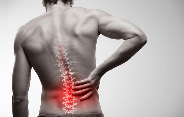
How does a lumbosacral hernia show?
A herniated disc can manifest itself with the following symptoms:
- Lumbar pain.
- Pain when walking that radiates to the thigh region.
- Numbness of the feet, fingers, areas on the surface of the lower leg and thigh.
- Heaviness in the legs.
- Stiffness of movement.
In order not to seek medical help too late, it is worthwhile to analyze the symptoms of the disease in more detail. They can be divided into three groups.
Pain syndrome
Lumbar spine hernia pain is a key symptom. Already in the first stage pain occurs in the area of the damaged intervertebral disc, especially after an injury. They can gain or lose weight and then reappear. More often, the sacral region doesn't even hurt, but hurts, especially during physical exertion or prolonged sedentary work. When a person lies on a healthy side and flexes the leg, the pain subsides completely. This condition can last for several months.
With timely treatment for medical help, getting rid of the problem is easy. It is enough to say goodbye to bad habits and do the physical therapy exercises recommended by your doctor.
Every day the affected area will increase and the condition of the disc tissue will worsen. The transition to the second degree of the disease is signaled by increased pain. It can now not only be felt in the sacral area, but covers the entire lower back, radiates into the neck area, onto every spinal column muscle, buttocks, thighs, legs, feet and toes. Discomfort is manifested with physical activity, even if it is insignificant - coughing or sneezing.
Vertebral syndrome
Increased pain in the second stage is accompanied by constant spasms of the back muscles. This leads to even greater discomfort for the patient. He cannot move freely, straighten his back, straighten himself. The gait of such a person becomes uncertain, he always leans on the side opposite the patient, sags.
A person's quality of life deteriorates due to impaired coordination of movements. He is not good at performing the tasks set at work, and active rest from constant pain becomes unrealistic.
Radicular Syndrome
If an inguinal hernia is left unattended by a doctor, the progressive disease leads to compression of the spinal roots, which causes them to die and blood access to the tissue of the damaged intervertebral disc becomes almost impossible. Symptoms characteristic of severe stages of the disease appear:
- Weakening of the leg muscles. The patient cannot squat, stretch, or jump. Climbing stairs is also difficult for him.
- Numbness of the affected area and surrounding areas. The skin becomes insensitive and pale, there are goose bumps and tingling sensations. Patients complain of hyperhidrosis in the affected area and legs or, conversely, excessive dryness of the skin.
- Lumbago. The patient suffers from lumbago in the lumbar region with acute, stabbing pain that increases with every movement. If left untreated, it leads to the destruction of the hip and knee joints.
- A noticeable thinning of the painful leg, which leads to postural asymmetry.
- Disruption of the pelvic organs. Urological and gynecological complaints are aggravated, libido disappears, diarrhea, urinary incontinence are possible.
In severe cases of spinal hernia, there is a risk of paralysis, disability, and even death.
Diagnosis of pathology
When a person has severe lower back pain, they need to make an appointment with a neurologist. He will conduct an examination with medical tests:
- Identification of reflexes from the tendons of the lower extremities.
- Test leg raises.
- Determination of heat or cold sensitivity, pain and vibration over the entire surface of the legs, thighs, buttocks, abdomen and back.
The doctor will then refer the patient for an MRI or CT scan of the lumbar spine. With the help of tomographic methods, a three-dimensional image of the affected area is made. It can be used to determine the location and size of the hernia, the stage of the disease.
If there is a risk of spinal cord injury, electromyography, neurography, and contrast agent myelography are also prescribed. Based on these studies, the doctor will determine if urgent surgery is needed.
Treatment of herniated discs
A vertebral fracture is treated both conservatively and surgically. The choice of technique depends on the stage of development of the disease, the presence of concomitant diseases and contraindications.
Conservative therapy
The therapeutic course is primarily aimed at relieving pain and relieving the patient's condition.
What drugs can a doctor prescribe:
- Drugs that relieve pain and inflammation. In case of an exacerbation - in the form of injections. When the acute pain is relieved (three to four days are usually enough), oral drugs with a similar effect are prescribed.
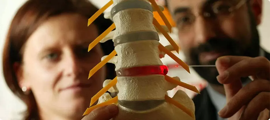
- Novocaine blockade with the addition of corticosteroids. A similar method can stop pain for two weeks at a time. Usually, a blockade is done with injections into different parts of the damaged disc.
- Centrally acting muscle relaxants. They reduce muscle activity by relieving pain cramps.
- Vitamin-mineral complexes with a focus on the elements of group B. They loosen the muscles slightly, help with tissue regeneration and the transmission of nerve impulses.
After relieving pain syndrome, the use of drugs decreases. Treatment of the disease is through physical therapy and physiotherapy.
Depending on the patient's condition, physiotherapeutic methods of treatment are also selected. It can be:
- Treatment with heat or electric shock.
- Electrophoresis with anti-inflammatory drugs.
- Acupuncture and acupressure.
- Hirudotherapy.
- Hydromassage.
Normal massage is allowed only if there is no pain syndrome. A more effective physiotherapy treatment is manual therapy with post-isometric relaxation.
Doctors strongly recommend that smoking patients give up cigarettes.
Diet adjustments are also important, especially for overweight patients. Fatty, savory foods, sweets and alcohol must be excluded from the menu. Eating a frugal diet with lots of vegetables and fermented dairy products will help the body endure the treatment better and shed the pounds on your back.
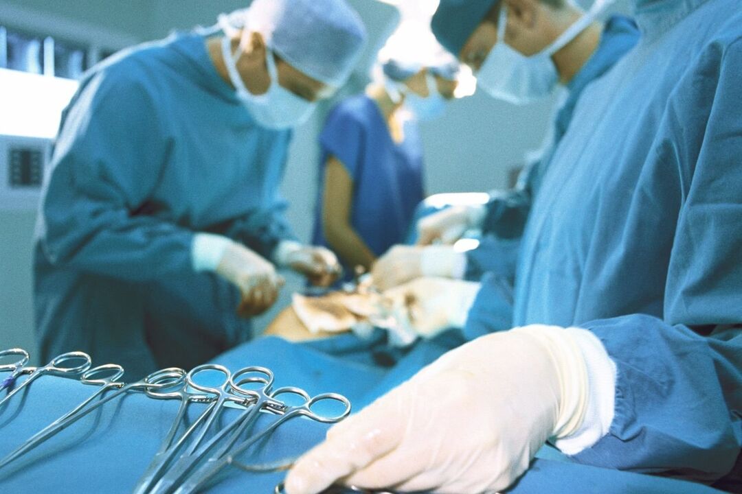
Surgical intervention
Conservative treatment usually lasts about two months. If it does not lead to the desired result, a decision is made to change therapeutic tactics or to perform an operation. The latter is prescribed for severe pain, loss of sensation in the legs, dysfunction of the pelvic organs. Depending on the complexity of the situation, the operation is carried out in the following ways:
- Endoscopic method. Three microincisions are made in the affected area. A camera is inserted into one for transmission to the monitor. The other two are used to remove the protrusion of the hernia using miniature instruments.
- By the method of percutaneous discectomy. The damaged core is removed through a puncture in the intervertebral disc and replaced with an artificial substance.
- By laser reconstruction. It is carried out in the form of punctures with a special needle, without dissecting tissue. Laser radiation heats the intervertebral disc structures and stimulates cell regeneration, as well as relieving pain.
In difficult cases, an endoprosthesis of the intervertebral discs is possible - the replacement of the injured organ with an implant.
Rehabilitation is required after complex surgical interventions. The operated person has to wear a corset and cannot take a seated position for about three months. The further rehabilitation phase includes the practice of therapeutic gymnastics and physiotherapy.
Preventive Techniques
Like any other disease, a herniated disc is easier to prevent than cure. What you need to do to keep your intervertebral discs healthy:
- Accurately calculate the loads if your job is related or if you are a professional athlete.
- Correct body weight (its index should not exceed 30).
- Choose a good mattress to sleep in the correct position (preferably on your back).
- Participate in gentle physical education, swimming, fitness.
- Incorporate exercises into the morning exercise to strengthen the muscle corset of the spine.
- Refrain from cigarettes.
- Eat well.
If following these rules becomes a habit, you run the risk of having a herniated disc just by accident.
A herniated disc is dangerous and has serious consequences, and advanced cases take a long time to treat. To avoid operations and complications, if you experience painful sensations in your back, you should see a neurologist.
Osteochondrosis
The term osteochondrosis itself is derived from two words: osteo - bone and chondru - cartilage. Put simply, it is an ossification of cartilage. Although this interpretation is fundamentally wrong. Some go even further in their delusions, believing that osteochondrosis is the build-up of salts in the joints. In addition, table salt should be eaten in large quantities.
Pathogenesis
In reality, everything works a little differently. And harder. And table salt, if it plays a role in causing osteochondrosis, is very indirect. Osteochondrosis is based on dystrophy and degeneration of the articular cartilage. This is not a disease in its own right, but a pathological process that can be observed almost anywhere where there is connective cartilage tissue.
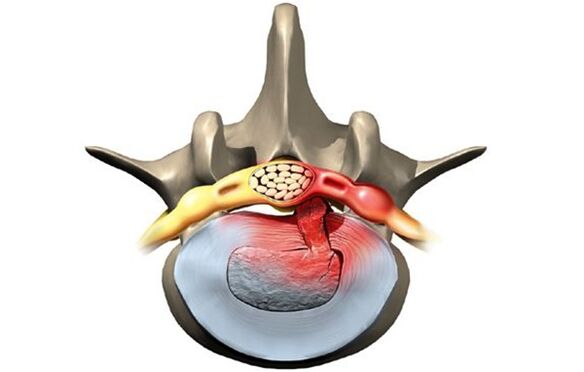
Still, osteochondrosis, overwhelmingly, affects the spine. Why so? The fact is that between the vertebrae there is a kind of spacer - intervertebral discs (intervertebral discs). The physiological task of these intervertebral discs is to cushion the vertebral bodies and to protect them from premature wear due to mechanical stress. The intervertebral disc consists of an inner liquid nucleus pulposus surrounded by an annulus fibrosus and an upper and lower endplate.
The intervertebral disc is subject to enormous mechanical stress, which leads to permanent damage to its structures at the cellular level. In humans, these processes are too pronounced - that is our reward for walking upright. To prevent the disk from being completely "erased", it must be constantly regenerated, ie restored. It is the balance of damage regeneration processes that determines the normal structure of the intervertebral disc. Another curious detail is that the intervertebral discs are not supplied with blood and nutrients via the blood vessels that have grown together in childhood, but rather diffusely from the bone tissue of the vertebral bodies. Again, paying for the ability to walk on two limbs, not four.
As a result, the intervertebral discs are easily injured in anatomical and physiological terms. Any negative process in the body leads to an imbalance in damage regeneration and the development of dystrophy and degeneration of the intervertebral discs. A structurally defective pane can no longer withstand the correct mechanical stress. Under excessive pressure from the vertebrae above, the intervertebral discs are shifted in different directions, usually to the side and back. This process is known as a herniated disc.
The bone tissue of the vertebrae that has lost its cartilage lining is also subject to mechanical wear. Due to the constant trauma on the anterior marginal surface of the vertebral bodies, pathological bone growths - osteophytes - are formed. Spondylosis develops. Due to the degeneration and displacement of the intervertebral disc, the intervertebral spaces shrink, the spinal canal narrows and the roots of the spinal nerves become so-called foraminal holes.
causes
The causes or etiological factors of osteochondrosis are diverse. They can be both local, i. H. caused by the pathology of the spine itself and general disorders at the level of the organism. Any pathology that leads to a violation of the structure of the spine or metabolic disorders can be considered the cause of osteochondrosis. In this regard there are:
- Changes in the configuration of the spine (scoliosis, pathological lordosis, or kyphosis)
- Other musculoskeletal defects - flat feet, narrow shoulder girdle, pelvic anomalies
- Spinal injury
- Weak immunity
- Metabolic disorders - osteoporosis, obesity, diabetes mellitus, thyroid disease
- Diseases of the cardiovascular system - atherosclerosis, hypertension
- Digestive disorders resulting in insufficient absorption of nutrients from the gastrointestinal tract
- Inheritance.
It should be noted that the above pathological conditions do not necessarily lead to osteochondrosis. This requires constant exposure to certain predisposing factors - hypothermia, malnutrition, sedentary lifestyle or, on the contrary, excessive physical exertion.
Symptoms
Osteochondrosis itself is an asymptomatic process. At the same time, the signs of intervertebral disc degeneration are diverse. How come? The fact is that the clinical manifestations of osteochondrosis are due to its complications - disc hernias, spondylosis, sciatica, narrowing of the spinal canal.
In addition, the clinic is very variable depending on the prevailing localization of the process in the cervical, thoracic or lumbar spine. The last section is most often affected as the lower back receives the maximum amount of physical activity. Signs of osteochondrosis of the lumbosacral region:
- Pain (lumbodynia, lumbago, sciatica)
- Restricted movement of the lower back and lower extremities (intermittent claudication)
- Here sensory disorders of the type of paresthesia - numbness, burning, crawling
- Pathological tension in the lumbar muscles
- Without treatment, functional disorders of the pelvic organs.
Cervical osteochondrosis is observed somewhat less frequently than lumbosacral. However, this pathology is also quite common. In addition to the typical pain symptoms (cervicalgia), reduced sensitivity and movements of the upper extremities, cervical osteochondrosis has its own peculiarities due to impaired blood flow to the brain. These properties manifest themselves:
- insomnia
- Headache, dizziness
- Periodic nausea
- General weakness, easy fatigue
- Fluctuations in blood pressure
- Occasionally toothache
- Behavioral reactions in the form of tears, irritability.
The chest area with osteochondrosis is relatively rarely affected. Patients in this case are people who are professionally forced to sit in a stable uncomfortable position - students, pupils, programmers, office workers. The symptoms of osteochondrosis in this case are as follows:
- Chest pain and paresthesia
- Dyspnoea
- Feeling of heartbeat
- Restricted movement of the thoracic spine.
diagnosis
From all of the above, it is clear that osteochondrosis is a chameleon disease. Due to the similarity of the signs, it is easy to confuse it with cerebrovascular accident, hypertension, myocardial infarction, angina pectoris, neurotic disorders. Therefore, in order to make the correct diagnosis, a comprehensive complex diagnosis is required in order to correctly determine the symptoms and treatment of osteochondrosis.
In addition to the classic questioning and clarification of patient complaints, this diagnosis should include a medical examination and special examination methods. These methods include x-ray of the spine, ultrasound of the internal organs. Recently, computed tomography and magnetic resonance imaging have been used successfully to diagnose osteochondrosis.
treatment
Therapeutic tactics for osteochondrosis include the use of:
- Medication
- massage
- Physiotherapy procedure
- Physiotherapy (movement therapy)
- Manual therapy
- Acupuncture.
Medicines for osteochondrosis are mainly aimed at relieving pain and eliminating inflammatory processes in the nerve roots. NSAID drugs are used for this purpose. In various combinations, these drugs are often used in the form of ointments, injections and tablets for the treatment of osteochondrosis. It should be remembered that these drugs have a negative effect on the liver, stomach and intestines. As a result, they can aggravate metabolic disorders in osteochondrosis. They relieve the pain of the blockage well with local anesthetics. The effect of these agents is short-lived and in no way affects the course of osteochondrosis as a whole.
With the help of drugs such as chondroprotectors, immunostimulants and vitamins with minerals, it is possible to improve metabolic processes at the local and body level. Chondroprotectors are used in tablets, ointments and ampoules. Among the tonics, vitamin C, group B, is used in combination with minerals. In this regard, calcium supplements are most preferred. In fact, contrary to some erroneous claims, osteochondrosis is not based on an excess, but only on a lack of calcium.
After successful exacerbation relief, physical therapy, massage, and exercise therapy will be shown. The physical methods used are electrophoresis with calcium, phonophoresis with hydrocortisone, amplipulse, and paraffin therapy. All of these measures are aimed at eliminating pain and inflammation in the nerve roots, ligaments, and muscles. Massage for osteochondrosis is carried out according to the generally accepted method. The massage zone is selected depending on the location of the osteochondrosis. The expansion of the range of motion is achieved with the help of exercise therapy. At the beginning, in the exacerbation phase, there are practically no dynamic loads. The patient is always in an optimal posture. At this time, it is desirable to wear immobilization devices - a lumbar corset, Shants' neck collar. When the exacerbation is eliminated, the volume and duration of movements during exercise therapy increase.
Recently, in the treatment of osteochondrosis, non-traditional methods of treatment have been used - acupuncture, manual therapy, osteopathy. Acupuncture is an effect on specific biologically active points along the spine, on the auricles, on the hands and on the feet. In manual therapy, the normal position of the vertebrae and intervertebral discs is restored through manual intervention by the hands of a specialist. And in the course of osteopathy, targeted techniques ensure the structural integrity of the musculoskeletal system. In the absence of the effect of conservative measures to treat osteochondrosis, persistent pain, complications, surgery is indicated. The pathologically displaced intervertebral disc is removed. A microdiscectomy is currently being performed for this purpose - endoscopic removal of a displaced intervertebral disc.